Learn

Skeletal Muscle Structure
Understanding muscle structure is important to the understanding of muscle contraction. Image 1 illustrates the structures making up skeletal muscle. Take a moment to look at the image. We will learn about each structure.
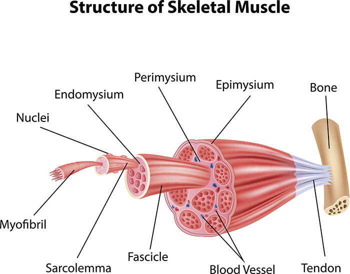
Image 1: Structure of Skeletal Muscle
Open larger version of Image 1 here.
There are 3 major types of connective tissue found within the skeletal muscle:
- Epimysium – is a connective tissue covering the entire muscle.
- Perimysium – is a connective tissue covering a group of muscle fibers.
- Endomysium – is a connective tissue covering individual muscle fibers.
Find these on the image above.
- Fascicle – is another name for a group of muscle fibers.
- Sarcolemma – is the cell membrane of a muscle fiber.
- Fiber – is a single muscle cell.
- Myofibrils – are the contractile structures of the cells made up of repeating protein units called sarcomeres.
- Myofilaments – are the filaments of myofibrils constructed from the proteins actin and myosin. (These myofilaments are illustrated in the image protruding from the myofibril and not labeled.)
This is a concept map of the skeletal muscle organization from the macroscopic level to the microscopic level:
Image 2: Skeletal Muscle Organization
Open larger version of Image 2 here.
Go to Get Body Smart: Skeletal Muscle Fiber Location and Composition for an interactive diagram of these structures. Be sure to click the highlighted words on that page.
How a Muscle Contracts
To understand how a muscle contracts, let's look at the structure of a sarcomere below. Sarcomeres are the functional contractile units of muscle fibers that are made up of thick (myosin) and thin (actin) filaments.
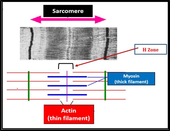
Image 3: Sarcomere Structure
Open larger version of Image 3: Sarcomere Structure.
- Notice these filaments illustrated in red and blue on the bottom part of the Image 3: Sarcomere Structure. The top part of the image is a stained microscopic view of these filaments.
- The H Zone is the area between the actin filaments. This zone will narrow as the muscle contracts due to myosin arms pulling the actin closer together.
On Image 4 below, notice the narrow H Zoneof the sarcomere in the image on the right. This happens when the muscle contracts after receiving a signal from a neuron. The neuron releases acetylcholine a neurotransmitter stimulating muscle contractions which is taken up by the muscle resulting in a cascade of events. These events culminate in contraction and relaxation of the muscle.
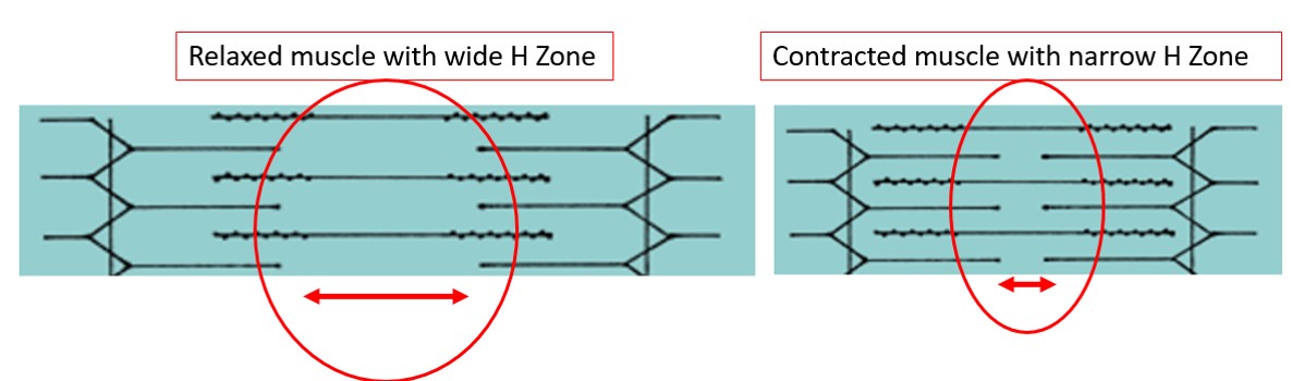
Image 4: H Zone diagrams
Open larger version of Image 4: H Zone diagrams.
Summary
Summary of the normal events in muscle contraction:
- A neuron sends a signal (action potential) to the muscle fibers.
- The neuron releases acetylcholine.
- Acetylcholine causes an action potential along the surface of the muscle.
- The action potentially causes calcium ions to be released from the sarcoplasmic reticulum (an organelle in the muscle).
- Calcium binds to a protein causing myosin and actin to connect (cross ridge formation).
- Cross bridge cycling results in muscle shortening as long as ATP is available.
Summary of the normal events in muscle relaxation:
- Calcium ions are taken back to the sarcoplasmic reticulum.
- Calsequestrin binds and stores calcium in the sarcoplasmic reticulum.
- Actin and myosin cannot bind anymore without calcium.
- Sodium (Na) is pumped out of the cell in exchange for potassium (K).